How can you suffer from hypertension at a young age?The cause is in this chest tablet ...
Author:Cardiovascular Channel in the Time:2022.08.17
*For medical professionals for reading reference

Do you think of this cause?
Point of learning
In this case, young women patients were found to be hypertension. When excluding secondary hypertension, the upper left hypertrophy was widening, and further examination was diagnosed as aortic contraction and tumor. Hypertension is caused by aortic contraction. The blood pressure in the cavity is returned to normal after treatment.
Young women, the unit's physical examination found hypertension, and the upper limb sleeve band blood pressure was around 160/80mmHg.
Common risk factors for young primary hypertension include: family history, obesity, hyperlipidemia, bad life hobbies (smoking, drinking), mental tension, anxiety, and so on.
Except for a little obesity (BMI 25) in this case, there are no other risk factors. The patient took the antihypertensive medicine for a while, but the effect was not good. The high voltage was about 140mmHg. I thought that I would take antihypertensive drugs when I was so young. I still went to the hospital for examination.
As a result, the patient went to the hospital for a series of examinations to exclude whether it was secondary hypertension. In the examination of common secondary hypertension, a chest tablet attracted clinical attention (Figure 1).
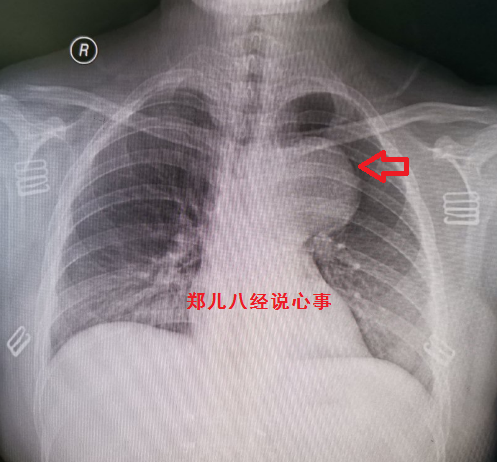
Figure 1 The chest sashimi shows the wide vertical shadow increase (shown in the red arrow)
Further physical examination found that heart murmurs merge the heart disease? Or functional noise? Cardiac ultrasound examination was excluded (Figure 2).
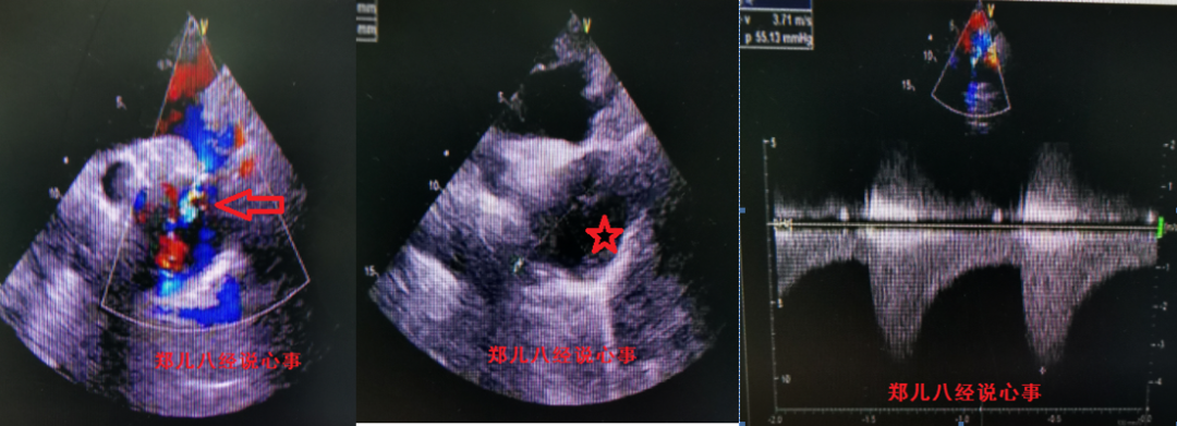
Figure 2 Cardiac ultrasound shows the stenosis of the starting part of the aortic arch of the aorta, 6mm in diameter of the narrowest part (shown in red arrows). After narrowing the anotal artery expansion, the aneurysm formation, 73x53mm in diameter (shown in the Red Star). The deviation difference between the aortic narrowing is 55mmHg. No other internal deformities were merged.
The upper septum wide range in the chest tablet is the aortic lowering the aorta in the color Doppler ultrasound, so the CTA examination is diagnosed (Figure 3 ~ 4).

Figure 3 axis CTA shows stenosis of the initial part of the aorta (maximum diameter 6.18mm, shown in red arrows). Considering the aorta narrowing, the red star shows the formation of post aneurysm, with a maximum diameter of 6.8cm.
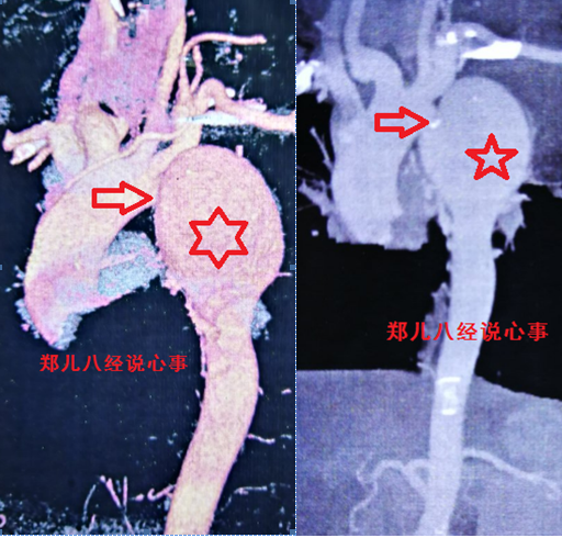
Figure 4 CTA three -dimensional reconstruction shows the formation of aortic narrowing and tumped aneurysm (shown in red arrows and Red Star)
At this point, the cause of the young female patient behind the hypertension is gradually clarified, which is secondary hypertension, aortic narrowing, and merging aortic tumor at the same time.
What is aortic narrowing
Aortic narrowing refers to congenital thoracic aortic limitation stenosis, the lumen is smaller or even blocked, and the blood flow is blocked. Aortic narrowing occurs near the arterial catheter or arterial ligament, but sometimes it can also occur at the proximal lower end of the left collarbone (Figure 5). Some people think that the arterial pipe fibrosis is affected during the process of the fibrosis of the arterial pipeline and the result of excessive narrowing of the aortic gorge or the aortic gorge.
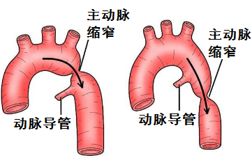
Figure 5 Aortic narrowing to form a schematic diagram
Clinical manifestations of aortic narrowing
The clinical symptoms of aortic narrowing are related to the narrowing site (anterior type of arterial duct, the posterior type of the arterial catheter), the degree of narrowing, the formation of the side branch, and whether the sideline formation is combined with other heart deformities. For those with mild degrees, there is a difference in high blood pressure, differences of upper and lower limbs, or heart murmurs.
For example, patients in these cases find hypertension, and the body is found in the physical examination, the heart of the heart is found, the blood pressure of the limbs is measured, the upper limbs are basically the same, and the blood pressure of the lower limbs is 50 ~ 70mmHg.
Treatment of aortic narrowing
The treatment plan for aortic narrowing mainly includes surgical treatment and intravus treatment. In this case, the patient refused to treat surgery, and conducted a series of intra -cavity feasibility assessments based on the specific condition of the patient (Figure 6-12).
(1) Circuit blood vessel evaluation;
(2) How to establish orbit from the aorta to the aorta through the narrowing site;
(3) Choose the type and size of the bracket;
(4) Is the anchoring area sufficient;
(5) The plan for emergencies during the intravusal treatment.
After sufficient preoperative evaluation, intra -cavity treatment was performed in the hybrid operating room.

Figure 6 before surgery CTA 3D reconstruction evaluation to enter the road blood vessels (shown in red arrows)

FIG. 7 The aortic photocopystics during the operation showed the bilateral iliac arteries (shown in the red arrow) and the formation of the aura pulse (shown in the Red Star).
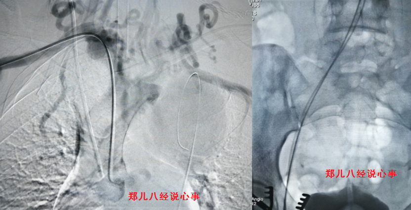
Figure 8 The right upper limb brachial artery road enters the single -curved pipe, and enters the aorta through the aortic narrowing site
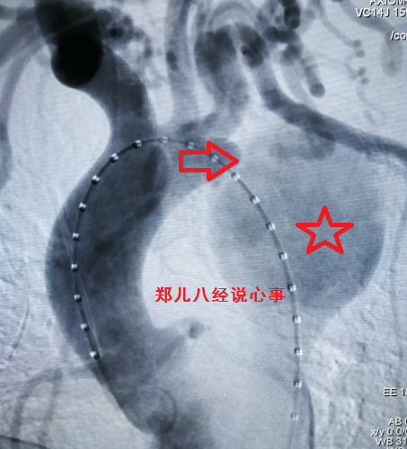
Figure 9 By exchanging the guide wire, the establishment of the femoral arterial path to the aortic orbit, the aortic photocopy of the aortic shadow shows the aortic narrowing and the aortic reducing aortic tumor (red arrows and red stars), and also show that the head and neck are more rough and large generations of large generations. Circulation of vascular side branches.

Figure 10 implant the bracket, the left cervical arteries open the window, cover the levelasmal artery opening, and the angiography shows that the left cervical arteries are unobstructed (the red arrows are shown in the red arrow). Show in the arrow)
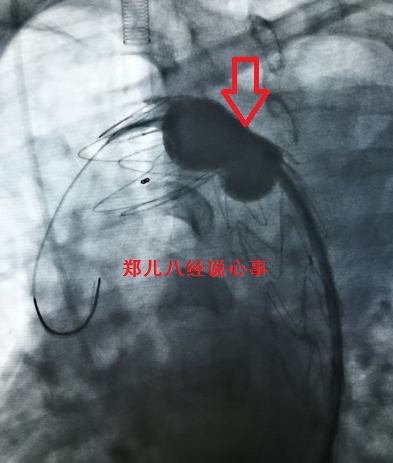
Figure 11 The narrowing part of the balloon expansion bracket

Figure 12 After the balloon is expanded, it will be taken again, the bracket is opened, and the left cervical artery is not visible (shown in the red arrow), the right axillary arterial arterial arterial arteries artificial blood vessels
The surgery was smooth, and the patient recovered well after surgery. The review of the CTA was reviewed as shown in Figure 13 ~ 14. After more than 4 years of follow -up, the blood pressure was normal, and the blood pressure on the upper and lower limbs returned to normal steps.

FIG. 13 Review the expansion of the aortic narrowing area (shown in the red arrow), and the aneurysm isolate (shown in the white arrow)
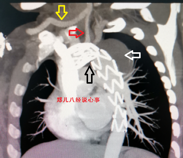
FIG. 14 Review the 3D reconstruction of the CTA showing the right axillary arteries -the left cervical artery artificial vascular is unobstructed (shown in the yellow arrow), the left cervical arteries are normal (shown in red arrows), and the aortic narrowing and expansion (shown in black arrows), Anesthesia isolated (shown in white arrows) This example summary:
(1) Patients with young patients should be measured for the first diagnosis of hypertension and compare bilateral upper and lower limb blood pressure;
(2) The aortic narrowing and seeing cardiovascular malformations are very important. Detailed physical examination is important;
(3) The treatment of aortic narrowing includes surgery and internal treatment;
(4) Aortic narrowing and combined with aortic tumor, internal treatment of the cavity should be evaluated in detail and fully planned.
More exciting cases
- END -
Open the list to gather the "unveil list of the list" in Qingdao High -tech Zone, the work ending fruit

Popular Network · Poster Journalist Jin Lixiu Correspondent Li Qingjian Qingdao r...
Ganzhou: The "old" residents enjoy the "new" life in the old community

Ganzhou Rong Media (Reporter Ren Ye) Ganzhou District has always adhered to the pe...