Laser provides new tools for the treatment of bone cancer.
Author:Science fiction Time:2022.09.20
Science Fiction Network September 20 (Wang Xiuxia) Due to technical reasons and other restrictions, doctors often cut off some healthy tissues when removing cancer cells. Recently, researchers at the California Institute of Technology have developed a new laser diagnostic imaging technology that can increase surgical cutting accuracy by 10 times, retain a healthy organization of 1,000 times more than before, and it is easier to recover after surgery.
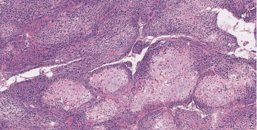
Among the many methods for treating cancer, the oldest and most respected methods are to remove all cancer tissues in the human body through surgery, while retaining the surrounding healthy substances as much as possible. However, it is difficult to clear the boundary between cancer tissue and health tissue. Especially when facing bone cancer, many health tissues are removed by accident, and the operation is extremely difficult. Scientists have been exploring the method of accurately identifying cancer cells.
Relying on dissolving is to determine whether a bone contains the most primitive traditional method of cancer cells. First of all, the bone block will be removed from the human body and then sent to the laboratory, where its hard calcium base quality will be slowly dissolved, leaving only live cells. Doctors will then perform sliced and imaging operations on the remaining materials. Because the process may take one to seven days to complete, it is very time -consuming. Doctors cannot rely on it to determine the health of the tumor and nearby bones during the operation. Therefore, they often cut off some bone tissues that should not be cut off. It is not good for patients' health.
In contrast, the new imaging technology developed by California Institute of Technology is better. This imaging technology is known as a real-time 3D contour scanning UV sound microscopy technology or UV-PAM, which aims to replace the traditional method of identifying cancerous bone tissue. The working principle of UV-PAM is to use laser to make the molecular vibration in the living tissue. These vibrations occur with ultrasonic frequencies, which can quickly imagine tissue and organs, just like watching the baby color Doppler ultrasound with ultrasonic waves. Since the process only takes a few minutes, it allows doctors to distinguish health bones from cancer bones in time during surgery.
Experts said: "The growth of bones is very difficult. If you cut off some healthy bones, it is difficult to grow back. Fortunately, we now have UV-PAM, which can provide imaging for doctors within 11 minutes. As a result, through images, doctors can use the knowledge learned to determine which are cancerous tissues and which are healthy tissues, so as to achieve accurate cutting. "
At present, this technology is proven only in the laboratory environment. Researchers say they want to bring this technology into the real world and clinical medicine. They will also formulate further improvement plans to provide more fine space resolution and higher imaging speed in order to see some details in the nucleus faster.
- END -
Baiyin Municipal County and District People's Society Director Live Bringing Goblies Warm Action Start

Reporter Wu XiaoIn order to implement the decision -making and deployment of the n...
Hebei: Tax and benefit to protect people's livelihood fine service for development

Since the implementation of the new combined tax support policy, the State Adminis...